Pancreatitis can present as sudden, severe epigastric pain or a subtler chronic illness, and distinguishing acute from chronic disease changes both tests and treatment. This guide explains how pancreatitis is diagnosed, when imaging or endoscopy are needed, the immediate hospital steps and longer term care, and the specific red flags that warrant surgical evaluation. For patients in Dhaka, it also outlines what to expect when consulting a pancreatic surgeon at Popular Medical College Hospital with Dr Murshidul Arefin.
1. Overview of pancreatitis and why it matters in Dhaka
Key point: In Dhaka, the clinical distinction between acute pancreatitis and chronic pancreatitis is the single most important decision point because it determines urgency, diagnostics, and whether surgery is likely to be needed.
How acute and chronic pancreatitis behave in real practice
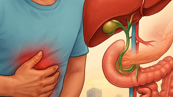
Acute pancreatitis usually presents with sudden severe upper abdominal pain, nausea, and elevated lipase. It ranges from mild illness that resolves with supportive care to severe disease with organ failure and pancreatic necrosis that requires ICU level care and possible delayed intervention. Chronic pancreatitis is progressive pain and malabsorption from long term pancreatic inflammation and scarring; treatment focuses on pain control, enzyme replacement, and targeted surgery for complications or intractable pain.
- Most common causes in Dhaka: gallstones and alcohol-related disease
- Other important causes: hypertriglyceridemia, medications, post-ERCP injury, autoimmune pancreatitis, and hereditary pancreatitis
- Less obvious contributors: obstructing tumors or biliary strictures which require imaging and surgical review
Practical insight: Transabdominal ultrasound is a workhorse in Dhaka for detecting gallstones and biliary dilation, but it misses small duct stones. When clinical suspicion remains high, MRCP or endoscopic ultrasound provide better ductal detail – accept the tradeoff of higher cost and scheduling delay for more reliable diagnosis. Contrast CT is best for assessing severity and complications but is most informative 48 to 72 hours after symptom onset.
Concrete Example: A patient arrives to a Dhaka emergency department with severe epigastric pain and lipase five times normal. Ultrasound shows gallstones without biliary dilation and no cholangitis. In practice this patient will get supportive care, early surgical review, and if stable should be offered laparoscopic cholecystectomy during the same admission to prevent recurrence. For duct stone suspicion, the team will arrange MRCP or coordinate ERCP with gastroenterology.
Early surgical involvement reduces repeat admissions for gallstone pancreatitis and shortens the time to definitive treatment.
lipase results to your surgical consultation at Popular Medical College Hospital for faster decision making. See cholecystectomy services for scheduling.Judgment: In Dhaka practice, assume gallstones or alcohol until proven otherwise. Do not rely solely on a normal ultrasound to exclude an obstructing stone if clinical signs point to biliary obstruction – escalate to MRCP or endoscopic ultrasound rather than delaying care. Next consideration: know the red flags that make pancreatitis an emergency.
2. Recognizing symptoms and red flag signs that need urgent care
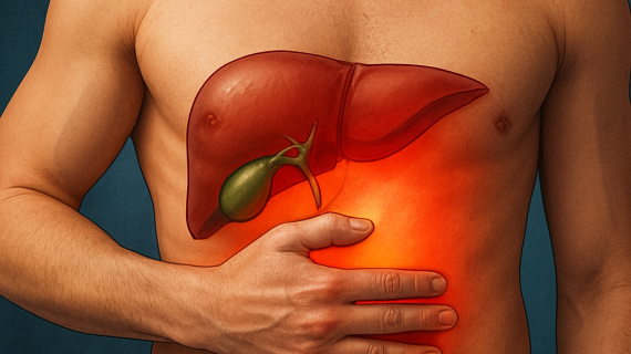
Immediate clinical reality: severe, constant upper abdominal pain that often radiates to the back is the most reliable early clue that pancreatic inflammation is present and needs assessment. Pain alone does not predict how sick someone will become, but its character and associated features determine whether you can wait for outpatient follow up or require urgent hospital care.
- Typical pancreatitis symptoms: severe epigastric pain (often radiating to the back), persistent nausea and vomiting, reduced appetite, abdominal bloating or distension
- Other common findings: fever, yellowing of the skin or eyes (jaundice), light-headedness from dehydration
- Red flags that need emergency assessment: fainting or persistent low blood pressure, trouble breathing or fast breathing, confusion or decreased alertness, little or no urine output for several hours, worsening or rapidly spreading abdominal tenderness (suggesting peritonitis), high fever or rigors, visible bruising around the umbilicus or flanks (Cullen or Turner signs)
- Specific concern for biliary obstruction or cholangitis: fever with jaundice and severe right-upper-quadrant pain — this combination needs urgent biliary drainage such as ERCP
Practical limitation to keep in mind: blood tests and early ultrasound can be misleading. Serum lipase is more sensitive than amylase but may be normal if tested very early; transabdominal ultrasound commonly misses small duct stones. That makes the history and appearance of red flags critical—do not rely solely on lab number or an initial normal scan to dismiss a worsening patient.
Concrete example: a 45-year-old woman in Dhaka with sudden epigastric pain and repeated vomiting felt somewhat better at home after analgesics, but developed fever and passed almost no urine over 12 hours. She was hypotensive on arrival and required aggressive IV fluids, ICU monitoring, and urgent imaging — a typical progression from what first seemed like a mild episode to severe pancreatitis with early organ dysfunction.
Judgment for patients and families: waiting out severe abdominal pain is a common mistake. Early hospital assessment reduces the chance of avoidable organ failure or infected pancreatic collections. If any red flag is present, present to the nearest emergency department rather than delaying for outpatient tests.
What to bring to expedite care in Dhaka: recent blood results (lipase/amylase, liver tests), any ultrasound or CT images on CD or digital copy, list of medications, and a short timeline of symptom onset and alcohol use or gallstone history. If you plan to see a specialist at Popular Medical College Hospital, bringing prior imaging shortens time to a definitive plan — see consultation. For general guidance on emergency signs see NIDDK pancreatitis resource.
3. Diagnostic tests and imaging pathway explained for patients
Direct point: the diagnostic pathway for pancreatitis separates two tasks: confirming pancreatic inflammation with blood tests and using imaging to find the cause and any complications. One without the other leaves unanswered questions that change treatment choices.
Initial blood tests and what they tell you
Core test: serum lipase is the preferred marker because it stays elevated longer and is more specific than amylase. Expect values to peak within the first 24 hours but know that very early testing can be falsely low.
Other useful labs: liver function tests, bilirubin, full blood count, C reactive protein, and triglycerides. These are not optional — they identify gallstone obstruction, infection risk, hypertriglyceridemia as a cause, and the inflammatory trend that predicts complications.
Imaging sequence and practical tradeoffs
First line: transabdominal ultrasound is quick, cheap, and widely available in Dhaka and it is best for gallstones and bile duct dilation. Limitation: it misses small common bile duct stones and can be degraded by bowel gas or obesity.
If ultrasound is inconclusive: choose between MRCP and endoscopic ultrasound. MRCP is noninvasive and excellent for anatomy; endoscopic ultrasound is more sensitive for tiny duct stones but requires sedation and an endoscopy team. The tradeoff is sensitivity versus invasiveness and cost.
When CT matters: contrast enhanced CT or CT pancreas is primarily for assessing complications such as fluid collections, necrosis, or pancreatic necrosis extent. In clinical practice CT is most informative after the first 48 to 72 hours when necrosis becomes apparent, but do not delay an urgent CT if the patient is deteriorating.
Role of ERCP: ERCP is a therapeutic procedure for removing duct stones or draining infected bile and is not the first diagnostic test. It carries a risk of causing pancreatitis, so it is reserved for clear indications such as cholangitis or persistent biliary obstruction.
Practical workflow in Dhaka: coordinate imaging with your surgical or gastroenterology team rather than ordering every test. Uncoordinated testing increases cost and delays the definitive step, whether that is ERCP, cholecystectomy, or conservative management.
Concrete example: a 52 year old man presents with typical pancreatitis and a nondiagnostic ultrasound. MRCP shows a small stone in the distal common bile duct. The team schedules ERCP the same day to remove the stone and then plans laparoscopic cholecystectomy within the admission to prevent recurrence.
Important: if blood tests, clinical course, and initial imaging disagree, trust the clinical trajectory. Escalate to MRCP, EUS, or urgent ERCP based on symptoms and signs, not just single abnormal results.
Judgment for patients: insist on targeted testing. Routine use of ERCP for diagnosis or immediate CT for every patient is overused and can harm or add cost. Ask your team why each test is recommended and how the result will change treatment.
4. Initial hospital management and monitoring for acute pancreatitis
Immediate priorities: start with hemodynamic support, effective pain control, and rapid risk stratification. These three actions determine whether care stays on a general ward or escalates to ICU and which interventions will follow.
Resuscitation: fluids, monitoring targets, and a practical tradeoff
Fluid strategy: use balanced crystalloid resuscitation guided by response, not a fixed formula. Begin with a 10 to 20 ml/kg bolus for patients who are hypotensive or clinically dehydrated, then reassess. Aim to restore perfusion while avoiding fluid overload, which worsens respiratory failure and abdominal compartment syndrome in practice.
| Parameter | Ward target (typical) | Why it matters |
|---|---|---|
| Mean arterial pressure | >= 65 mmHg | Ensures organ perfusion |
| Urine output | >= 0.5 ml/kg/hr | Easy bedside perfusion marker |
| Respiratory status | No tachypnea, SpO2 >= 94% on oxygen as required | Detects fluid overload and early lung injury |
| Heart rate | < 100 bpm (trend matters) | Persistent tachycardia suggests under-resuscitation or sepsis |
Practical monitoring note: place a urinary catheter early if urine output is unreliable. Bedside ultrasound for IVC variability or lung B lines helps refine fluid decisions and avoids unnecessary central lines in many patients.
Analgesia, organ support, and when to transfer to ICU
Analgesia approach: use titratable IV opioids as first-line for severe pain; patient-controlled analgesia is often preferable on the ward. Consider epidural analgesia in select patients with uncontrolled pain and no coagulopathy, but weigh the resource and monitoring needs.
ICU triggers: escalating vasopressor requirement, persistent hypoxia requiring >40 percent oxygen, oliguria despite fluids, altered mental status, or multi-organ dysfunction. Transfer early; delaying ICU transfer because a patient looks stable at one time point is a common cause of preventable deterioration.
Nutrition and infection: what changes practice
Nutrition: start enteral feeding within 24 to 48 hours when feasible because it reduces infectious complications compared with parenteral nutrition. Use a nasojejunal tube only if gastric feeding is not tolerated. Total parenteral nutrition is a last resort for patients with prolonged ileus or failed enteral feeding.
Antibiotics and infected collections: do not use antibiotics routinely for sterile pancreatic necrosis. Suspect infection with persistent fever, rising white cell count, or gas in a collection on imaging; then obtain cultures and plan drainage combined with targeted antibiotics rather than blind broad spectrum prophylaxis.
Intervention timing judgment: avoid early debridement of necrosis. In practice, most infected collections are managed with percutaneous or endoscopic drainage first and delayed necrosectomy only if the patient does not improve. This step-up approach lowers morbidity compared with immediate open surgery.
Concrete example: a 58 year old man arrives hypotensive and tachycardic after two days of vomiting. He received a 15 ml/kg crystalloid bolus, had a urinary catheter inserted, and urine output rose to 1 ml/kg/hr; analgesia was started with IV morphine PCA. On day 7 he developed fever and a tender collection on CT with gas; the team placed a percutaneous drain and started culture-directed antibiotics, avoiding early open necrosectomy.
Important: balance aggressive early resuscitation against the risk of fluid overload. Use clinical trends and bedside tools to titrate fluids rather than fixed high-volume protocols.
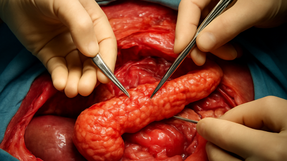
5. Endoscopic and surgical treatment options and when each is appropriate
Bottom line: use the least invasive effective approach first, but choose the modality that solves the problem rather than the one that is most convenient. Endoscopy removes stones and drains collections with lower short-term morbidity; formal surgery is reserved for structural disease, failed minimally invasive therapy, or oncologic resection.
Endoscopic options and realistic limits
ERCP (endoscopic retrograde cholangiopancreatography): best for clear biliary obstruction or cholangitis. Timing matters — urgent ERCP when fever, jaundice, and deranged liver tests point to retained duct stones; delaying in that scenario increases sepsis risk. ERCP is not a fix for all pancreatitis and can itself cause pancreatitis if used indiscriminately.
Endoscopic transmural drainage and endoscopic necrosectomy: effective for well-formed walled-off pancreatic collections that abut the stomach or duodenum. The advantage is lower physiologic stress compared with open debridement; the limitation is that collections must be accessible endoscopically and multiple sessions are often required. If your team lacks endoscopic ultrasound expertise, percutaneous or surgical options become necessary.
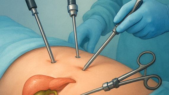
Minimally invasive and percutaneous approaches
Percutaneous drainage: often the quickest option for infected collections when access is safe. It stabilizes the patient and can be definitive for simple abscesses. Tradeoff: percutaneous drains may not clear dense necrosis and can create external fistulae requiring further management.
VARD and laparoscopic/minimally invasive necrosectomy: these techniques sit between percutaneous drainage and open surgery. They reduce pulmonary and wound complications compared with open necrosectomy but demand experienced teams and ICU support. In Dhaka practice, picking the right center matters — incomplete debridement or premature open surgery increases morbidity.
When formal surgery is the correct choice
Laparoscopic cholecystectomy: indicated to prevent recurrence after gallstone pancreatitis once the patient is clinically stable. Early cholecystectomy during the same admission is preferred when feasible, but hemodynamic instability or ongoing organ failure requires postponement.
Resectional surgery (distal pancreatectomy, pancreaticoduodenectomy/Whipple): reserved for neoplasms or select chronic pancreatitis cases with localized disease. These are major operations with significant long-term consequences such as endocrine insufficiency; decide only after staging, biopsy when needed, and multidisciplinary discussion.
Open necrosectomy: now a last resort when minimally invasive steps fail or immediate control of ongoing sepsis/hemorrhage is required. Rushing to open surgery before necrosis has demarcated or before attempts at drainage often increases bleeding and mortality.
- Decision factors surgeons weigh: clinical stability, infection status of collections, anatomy of the collection, local expertise (ERCP/EUS/advanced endoscopy), and goals of care.
- Local capacity matters: a center with experienced endoscopists and interventional radiologists can avoid many open procedures; if those services are unavailable, earlier surgical referral is appropriate.
Concrete example: A patient with gallstone pancreatitis and persistent cholestatic jaundice had same-admission ERCP with stone extraction followed by laparoscopic cholecystectomy two days later — this prevented a readmission. Another patient developed infected walled-off necrosis at week 3; the team placed endoscopic stents and performed serial endoscopic necrosectomies over three sessions, avoiding open debridement and shortening ICU stay.
Important: for necrotic collections, delaying definitive debridement until the collection matures (usually >3–4 weeks) when possible reduces bleeding and increases the chance that minimally invasive methods will succeed.
Judgment: choose the approach that clears the cause and minimises additional harm. Endoscopy and percutaneous drainage have shifted practice away from early open surgery, but those techniques require local expertise and multiple sessions. When a pancreatic mass is suspected, expedite staging and a surgical oncology review rather than attempting blind drainage or delayed resection without proper planning. For appointments, bring recent imaging and labs to speed multidisciplinary decisions — see consultation.
6. When to consult a pancreatic surgeon in Dhaka and what to expect at the first visit
Practical rule: see a pancreatic surgeon early when a scan or clinical course suggests structural disease, persistent organ dysfunction, or complications that are unlikely to resolve with medical care alone. Early surgical assessment does not mean immediate operation — it means a timely plan that coordinates ERCP, interventional radiology, endoscopy, or staged surgery as needed.
| Referral reason | Why a surgeon matters | Typical timing in Dhaka |
|---|---|---|
| Suspected pancreatic mass on CT/MRCP | Surgical staging, biopsy planning, and discussion of resection if appropriate | Urgent — appointment within days to 2 weeks; MDT staging promptly |
| Infected pancreatic necrosis or worsening sepsis with collections | Decide drainage strategy (percutaneous, endoscopic, or necrosectomy) | Immediate surgical review with interventional radiology/endoscopy coordination |
| Recurrent gallstone pancreatitis | Plan definitive biliary surgery (laparoscopic cholecystectomy) to prevent recurrence | Within the same admission if stable, or early outpatient scheduling |
| Symptomatic pancreatic pseudocyst or enlarging walled-off necrosis | Choose endoscopic or surgical drainage and discuss timing | Planned intervention after multidisciplinary review (weeks if not infected) |
| Persistent biliary obstruction or cholangitis | Coordinate urgent ERCP and potential follow-up surgery | Same-day to 48 hours depending on stability |
Preparing for the appointment
Bring the clinical dossier: digital imaging in DICOM format or CDs, written radiology reports, discharge summaries, operation notes, endoscopy (ERCP/EUS) reports, recent blood tests (lipase, LFTs, CRP, triglycerides), current medication list including enzyme supplements and diabetes therapy, and any pathology reports. Having these files ahead of the visit shortens decision time and avoids repeat testing.
- Imaging: DICOM MRCP/CT files or recent ultrasound
- Procedures: ERCP/EUS reports and fluoroscopy images if available
- Labs: recent serial lipase/amylase, LFTs, and triglycerides
- Records: discharge summaries and prior operative notes
- Medications: full list including pancreatic enzymes and insulin
- Administrative: ID, referral letter, and insurance/consent paperwork
What happens at the first visit
The first consultation is a focused clinical assessment and an image-led discussion. Expect a concise history emphasizing timing of pain, previous procedures (ERCP, stenting), alcohol use, and prior admissions. The surgeon will review the imaging with you using the original files, outline possible pathways (surveillance, ERCP, endoscopic drainage, percutaneous drainage, or planned surgery), and explain the immediate next steps and likely timeline.
Trade-off to understand: some problems are best fixed quickly (obstructing duct stones or cholangitis), others improve with conservative care and benefit from delayed intervention (mature necrosis or walled-off collections). In Dhaka the limiting factors are scheduling for EUS/ERCP, interventional radiology availability, and ICU capacity — these practical constraints shape the chosen plan as much as clinical indications do.
Concrete example: a 62-year-old man has a contrast CT showing a 3.5 cm lesion in the pancreatic body. At his first visit at Popular Medical College Hospital with Dr Murshidul Arefin, the surgeon reviews the CT and MRCP, arranges an endoscopic ultrasound-guided biopsy the same week, and places the case on the hepatobiliary multidisciplinary team agenda to decide between distal pancreatectomy and neoadjuvant oncologic treatment. The patient leaves with a clear diagnostic and scheduling plan rather than an immediate, unplanned operation.
Key point: if imaging suggests a mass or there is infected necrosis, do not delay referral — early surgical involvement shortens time to definitive diagnostics and reduces avoidable procedures.
7. Recovery, long term management, and preventing recurrence
Discharge expectations: after an acute episode most patients leave hospital once pain is controlled, oral intake is tolerated, and organ function has normalized. Expect a short course of analgesics, a clear plan for diet progression, and a clinic appointment within 1 to 2 weeks for early review. If you had necrotizing pancreatitis, anticipate a longer, staged follow up with imaging and possible interventions.
Practical components of recovery
Pancreatic enzyme replacement: patients with persistent steatorrhea, weight loss, or malabsorption usually benefit from enzyme supplements. Dosing is individualized; many people are undertreated in real practice because providers start low to save cost. Judgment: titrate to symptom response and weight gain rather than an arbitrary pill count.
- Nutrition strategy: progressive return to a normal diet focused on small frequent meals and a low-fat approach if steatorrhea is present; involve a dietitian for caloric targets and micronutrient checks.
- Diabetes and endocrine follow up: new or worsening hyperglycemia after pancreatitis requires testing and often joint care with endocrinology; pancreatic diabetes behaves differently and may need insulin earlier.
- Medication review: stop offending drugs if they were implicated, check triglycerides and arrange lipid-lowering therapy when hypertriglyceridemia was the cause.
Limitation and tradeoff: aggressive fat restriction can reduce symptoms but risks poor calorie intake and fat-soluble vitamin deficiencies. The practical balance is targeted fat moderation plus enzyme replacement rather than prolonged, strict low-fat diets that patients cannot sustain.
Preventing recurrence: remove the cause when possible. For gallstone pancreatitis definitive cholecystectomy prevents repeat attacks. For alcohol-induced disease meaningful risk reduction requires sustained abstinence and addiction support. For high triglycerides, expect medical therapy and diet counselling before you consider the episode controlled.
When recurrent pancreatitis needs specialist reassessment: two or more episodes, unexplained persistent pain, or new weight loss should trigger expedited review by a pancreatic specialist to reassess for obstructing lesions, ductal strictures, or genetic and autoimmune causes.
Concrete example: a 49 year old man discharged after moderate pancreatitis due to high triglycerides returns for follow up with ongoing loose stools and 4 kg weight loss. At Popular Medical College Hospital he started pancreatic enzyme replacement, met a dietitian for calorie dense, lower fat meals, and had a lipid clinic appointment to start fibrate therapy. Within 8 weeks his stools improved and weight stabilised.
Important: enzyme supplements treat malabsorption but do not reliably relieve pain from chronic pancreatitis; expect separate pain management planning if pain persists.
Next consideration: if you are in Dhaka and need coordinated follow up—imaging, dietetic support, or endocrine care—bring previous imaging and blood tests to your surgical or gastroenterology appointment and arrange multidisciplinary review at Popular Medical College Hospital via consultation.
8. How Dr Murshidul Arefin and Popular Medical College Hospital support patients with pancreatitis in Dhaka
Practical reality: Dr Murshidul Arefin leads a coordinated hepatobiliary-pancreatic service at Popular Medical College Hospital that pairs surgical decision-making with gastroenterology, interventional radiology, and critical care. The centre offers ERCP scheduling and stenting, endoscopic ultrasound (EUS) access, minimally invasive drainage and necrosectomy when indicated, laparoscopic biliary surgery, and staged pancreatic resections — all supported by on-site ICU for high-risk cases.
Important trade-off: advanced endoscopic and interventional radiology work best when scheduled as a team. In Dhaka the constraint is rarely the theory of care but the practical availability of EUS/ERCP slots and ICU beds. That means some patients are treated in a staged fashion: urgent life-saving drainage first, definitive endoscopic or surgical care once the patient is stable. Expect deliberate sequencing rather than immediate single-session fixes.
Typical patient pathway at Popular Medical College Hospital
- Triage and rapid review: acute patients get immediate resuscitation and a focused surgical assessment.
- Image-led planning: the team reviews CT/MRCP/EUS images together to decide ERCP, percutaneous drainage, endoscopic drainage, or surgery.
- Targeted intervention: perform the least invasive effective procedure first — ERCP for retained duct stones, percutaneous drain for accessible infected collections, or EUS-guided transgastric drainage for walled-off necrosis.
- Definitive care and timing: plan laparoscopic cholecystectomy during the same admission when safe, or schedule elective pancreatic resection after MDT staging for neoplastic disease.
- Coordinated follow-up: postoperative ICU support if needed, dietetic and endocrine referrals, and clear outpatient imaging and clinic timelines.
Concrete example: a 54-year-old with recurrent biliary-type pancreatitis was reviewed by Dr Arefin; MRCP suggested a distal bile duct stone not seen on ultrasound. The team performed ERCP with stone extraction on day 1, placed a temporary biliary stent, and discharged the patient for laparoscopic cholecystectomy two days later. This staged approach prevented another emergency readmission and compressed the overall treatment into one coordinated pathway.
Judgment: in Dhaka you gain the most by choosing a centre that manages the whole sequence — diagnosis, endoscopy, drainage, surgery, and ICU — rather than piecemeal referrals. Centres that cannot offer EUS-guided drainage or endoscopic necrosectomy will still treat pancreatitis, but patients face higher odds of open surgery or repeat procedures.
Bring your imaging files (DICOM or readable digital copies), recent blood tests, and a concise timeline of symptoms to the first visit to speed decision-making.
Next consideration: if you have recurrent attacks, a suspicious mass, or signs of infection around pancreatic collections, ask for an expedited surgical review. Early multidisciplinary triage at Popular Medical College Hospital changes both the immediate plan and the likelihood of avoiding major open surgery.