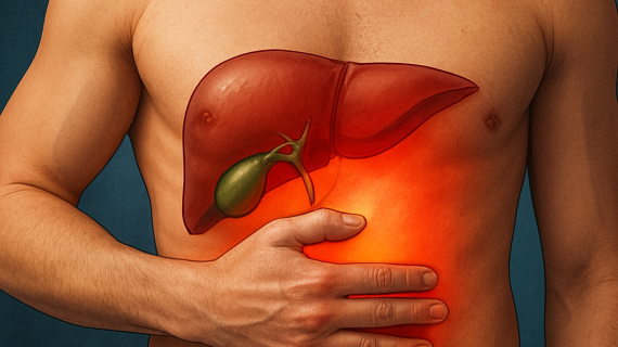
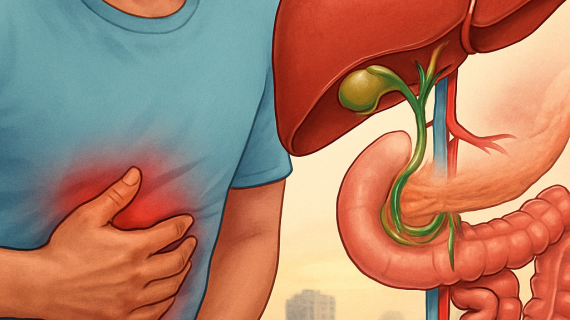
Liver cysts are a common finding in medical imaging, often discovered incidentally during investigations for unrelated conditions. While many are benign and require no intervention, a subset can present with complex characteristics, necessitating careful evaluation and management. Understanding the classification, imaging features, and available treatment options is paramount for clinicians to provide appropriate care for patients presenting with these lesions. This article aims to demystify the landscape of liver cysts, guiding you through their categorization, the diagnostic power of imaging modalities, and the spectrum of management strategies, from watchful waiting to surgical intervention.
The journey into understanding liver cysts begins with their classification. Like a diverse ecosystem, liver cysts can arise from various origins, each with distinct implications. Broadly, they can be categorized into congenital, traumatic, infectious, or neoplastic lesions. This foundational understanding allows clinicians to form initial hypotheses about the nature of a detected cyst.
Congenital Cysts
Congenital cysts are present at birth, developing due to malformations of the intrahepatic bile ducts during embryonic development. These are the most prevalent type of simple liver cyst.
Simple Hepatic Cysts
Simple hepatic cysts are fluid-filled sacs with thin, smooth walls and no internal septations or solid components. They are typically solitary but can be multiple and are often discovered incidentally. Their pathogenesis is thought to relate to dysgenetic bile ductules that become isolated and dilate.
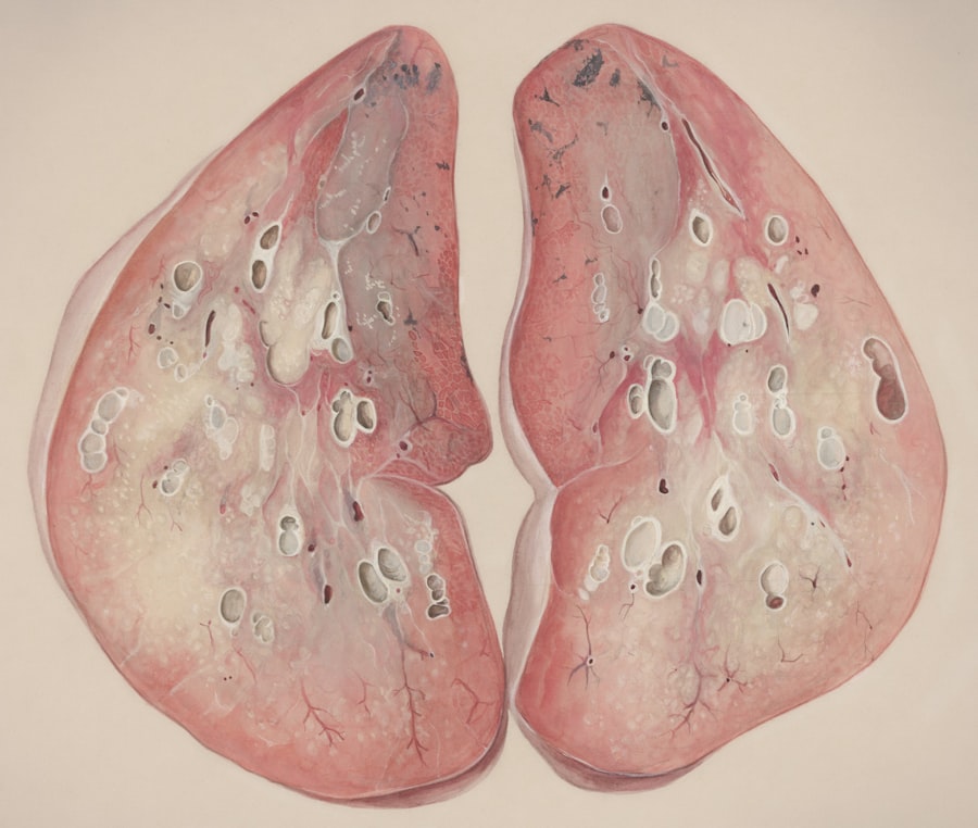
Polycystic Liver Disease
Polycystic liver disease (PLD) is a genetic disorder characterized by the development of numerous cysts throughout the liver parenchyma. It can be inherited as autosomal dominant (ADPLD) or autosomal recessive (ARPLD). ADPLD is more common and is often associated with autosomal dominant polycystic kidney disease (ADPKD), although liver cysts can occur in isolation. ARPLD is rarer and typically presents in infancy with severe liver dysfunction.
Traumatic Cysts
Traumatic cysts develop following injury to the liver. This can range from blunt abdominal trauma to penetrating injuries. The cyst may form as a result of blood or bile accumulation within the liver parenchyma, which then becomes encapsulated over time.
Hematomas and Bile Collections
Post-traumatic hematomas can organize and liquefy, forming cystic structures. Similarly, bile leaks following trauma can lead to the formation of biliomas, which are encapsulated collections of bile.
Infectious Cysts
Infectious etiologies are a significant consideration, particularly in endemic regions. These cysts arise from parasitic infestations or bacterial infections.
Echinococcal Cysts (Hydatid Cysts)
Echinococcal cysts are caused by infection with the larval stage of the tapeworm Echinococcus granulosus. These are a classic example of parasitic cysts and are often referred to as hydatid cysts. They have a characteristic multi-layered wall and can grow quite large. The World Health Organization (WHO) has developed a standardized classification for cystic echinococcosis, known as the WHO-Informal Working Group on Echinococcosis (WHO-IWGE) classification. This system categorizes cysts into five stages, from CE1 (unilocular cyst with a fluid anechoic content) to CE5 (calcified cyst), which helps in predicting infectivity and guiding treatment.
Pyogenic Liver Abscesses
While not true cysts in the typical sense, pyogenic liver abscesses can present as cystic lesions filled with pus. They are typically associated with bacterial infections and can be very serious. They are often multiloculated and contain debris.
Neoplastic Cysts
Neoplastic cysts represent a more concerning category as they can be premalignant or malignant. These arise from the proliferation of abnormal cells within the liver.
Cystadenomas
Hepatic cystadenomas are rare cystic neoplasms of the liver that arise from the biliary epithelium. They are considered premalignant lesions, with a reported risk of malignant transformation into cystadenocarcinomas in approximately 10% of cases. They are typically multiloculated and can contain mucinous fluid.
Cystadenocarcinomas
Cystadenocarcinomas are malignant neoplasms originating from the biliary epithelium, presenting as cystic masses with features of malignancy.
Other Neoplastic Cysts
Less common neoplastic cystic lesions include cystic forms of hepatocellular carcinoma and metastatic disease to the liver manifesting as cystic lesions.
In exploring the complexities of liver cysts, the article “Liver Cyst: Classification, Imaging-Based Diagnosis, and Management Options from Surveillance to Intervention” provides a comprehensive overview of the various types of liver cysts, their diagnostic imaging techniques, and the management strategies available. For those interested in enhancing their understanding of effective communication in medical writing, a related article on crafting engaging headlines can be found at Crafting Captivating Headlines. This resource offers valuable insights that can help medical professionals present their findings more effectively.
Imaging-Based Diagnosis
The advent of advanced imaging technologies has revolutionized the diagnosis and characterization of liver cysts. Medical imaging acts as the clinician’s eyes, allowing them to peer into the depths of the liver and differentiate between the many types of cystic lesions.
In exploring the complexities of liver cysts, it is beneficial to consider related research that delves into the nuances of liver health and disease management. A comprehensive article on this topic can be found at this link, which discusses various aspects of liver conditions, including diagnostic techniques and treatment options. Understanding these connections can enhance our approach to liver cysts, particularly in terms of effective surveillance and intervention strategies.
Ultrasound: The Initial Scout
Ultrasound (US) is invariably the first-line imaging modality for evaluating suspected liver pathology, including cysts. Its accessibility, cost-effectiveness, and lack of ionizing radiation make it an ideal screening tool.
Simple Cyst Appearance on Ultrasound
A simple liver cyst on ultrasound typically presents as a well-defined, anechoic (black) lesion with smooth, thin walls. Posterior acoustic enhancement, a bright echo behind the cyst, is a characteristic feature due to the homogeneous fluid density of the cyst, which allows sound waves to pass through with minimal attenuation. The internal echo pattern is uniformly anechoic, indicating fluid-filled contents. Doppler ultrasound demonstrates absent internal vascularity, further supporting a simple cystic nature.
Complex Cyst Features on Ultrasound
When a cyst deviates from the “simple” appearance, it is considered complex. Ultrasound can detect septations (internal divisions), internal echoes (suggesting debris, pus, or internal components), mural nodules (solid masses attached to the cyst wall), and calcifications. These features raise suspicion for more complicated diagnoses, including parasitic cysts, abscesses, or neoplastic lesions.
Computed Tomography (CT): Adding Detail
CT scans provide excellent cross-sectional anatomical detail and are invaluable in further characterizing liver cysts, particularly those identified as complex on ultrasound.
CT for Differentiation of Simple vs. Complex Cysts
On CT, simple cysts appear as well-defined, homogeneous, low-attenuation (dark) lesions with a smooth wall. Their attenuation values are typically close to that of water. Complex cysts can demonstrate a range of CT appearances. The presence of internal septations, calcifications within the wall or septa, irregular or thickened walls, and internal solid components are key indicators of complexity. CT is particularly adept at visualizing calcifications, which can be hallmarks of hydatid cysts or some neoplastic lesions.
Evaluating Cyst Contents and Borders
CT is superior to ultrasound in detecting subtle calcifications and assessing the relationship of a cyst to surrounding vascular and biliary structures. It can also help differentiate between fluid, necrosis, hemorrhage, and solid components within a cystic lesion based on attenuation values.
Magnetic Resonance Imaging (MRI): Unraveling Ambiguity
MRI is often the gold standard for characterizing indeterminate liver lesions, providing superior soft-tissue contrast compared to CT. It is particularly useful when ultrasound and CT findings are equivocal.
MRI Characterization of Cysts
On MRI, simple cysts demonstrate uniformly low signal intensity on T1-weighted images and very high signal intensity on T2-weighted images. This bright signal on T2 is a hallmark of fluid-filled structures. The cyst walls are typically thin and smooth with no enhancement after the administration of gadolinium-based contrast agents.
Advanced MRI Techniques
Diffusion-weighted imaging (DWI) can help distinguish between benign and malignant cystic lesions, as malignant lesions often exhibit restricted diffusion. Gadolinium-enhanced MRI is crucial for evaluating enhancement patterns within cyst walls or septations, which can indicate inflammation, vascularity, or neoplastic components. Specific sequences can also help assess the nature of cyst contents, such as the presence of proteinaceous material or blood products. MRI is also excellent for demonstrating the relationship of cystic lesions to the portal vein, hepatic veins, and biliary tree, which is critical for surgical planning.
Management Options: From Surveillance to Intervention

The management of liver cysts is a tailored approach, dictated by the cyst’s classification, imaging characteristics, and the presence or absence of symptoms. The spectrum of options ranges from a hands-off, watchful approach to active surgical intervention.
Surveillance for Simple Cysts
The vast majority of incidentally discovered liver cysts are simple cysts. Given their benign nature and very low risk of complications, these typically require only observation.
Indications for Surveillance
If a liver cyst meets all the criteria for a simple cyst on imaging – anechoic, well-defined, smooth thin wall, posterior acoustic enhancement on ultrasound, and no internal septations, solid components, or calcifications on CT/MRI – then routine follow-up imaging is usually not necessary. The prevalence of simple cysts in the general population is estimated to be between 2.5% and 5%, and they rarely cause symptoms or complications. Their stability over time further supports a conservative management strategy.
Long-Term Follow-Up Protocols
In some cases, particularly for larger simple cysts (e.g., >5 cm) or in patients with a history of other liver conditions, a single follow-up imaging study after 6-12 months may be considered to confirm stability. However, ongoing surveillance is generally not recommended as the risk of change is exceedingly low.
Management of Complex Cysts and Specific Etiologies
When a liver cyst exhibits features of complexity or is suspected to be of a specific etiology requiring treatment, a more active management strategy is warranted.
Management of Cystic Neoplasms
Cystic neoplasms, such as cystadenomas and cystadenocarcinomas, necessitate intervention due to their premalignant or malignant potential.
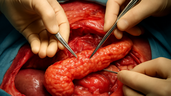
Surgical Excision
Surgical excision, typically via a hepatectomy or enucleation (removal of the cyst without surrounding liver tissue), is the treatment of choice for symptomatic cystic neoplasms or lesions with suspicious features. The goal is complete removal to prevent recurrence and malignant transformation.
Role of Liver Transplantation
In rare cases of diffuse polycystic liver disease with severe liver dysfunction and liver failure, liver transplantation may be considered as a life-saving intervention. This is usually reserved for patients who are otherwise good candidates for transplantation and have no significant extrahepatic manifestations.
Management of Hydatid Cysts
Hydatid cysts, caused by Echinococcus infection, require specific treatment protocols to eradicate the parasite and prevent complications such as anaphylaxis or cyst rupture.
Percutaneous Drainage and Aspiration
The European Association for the Study of the Liver (EASL) and the American Association for the Study of Liver Diseases (AASLD) recommend a combination of percutaneous drainage, aspiration, injection of a scolicidal agent (e.g., hypertonic saline or ethanol), and re-aspiration (PAIR technique) for uncomplicated hydatid cysts, particularly for CE1 and CE2 cysts. This approach aims to inactivate the parasite and drain the cyst contents.
Antimicrobial Therapy
Medical treatment with antiparasitic agents, such as albendazole or mebendazole, is often used in conjunction with or as an alternative to surgical or percutaneous interventions, especially for inoperable cysts or as adjunctive therapy to reduce the risk of recurrence.
Surgical Intervention for Hydatid Cysts
Surgical intervention, including cystectomy (removal of the cyst) and hepatic resection, may be indicated for complicated hydatid cysts, deeply embedded cysts, infected cysts, or when percutaneous treatment is not feasible. Meticulous drenching of the operative field with scolicidal agents is crucial to prevent spillage of protoscolices, which can lead to anaphylaxis or secondary hydatid disease.
Management of Infected Cysts and Abscesses
Liver abscesses, whether pyogenic or amoebic, typically require prompt drainage and antimicrobial therapy.
Percutaneous Drainage and Antibiotics
Percutaneous drainage of liver abscesses, guided by imaging, is often the initial treatment of choice. This allows for the removal of purulent material and subsequent irrigation. Broad-spectrum antibiotics are administered concurrently to combat the underlying bacterial infection.
Surgical Drainage
In cases of multiple abscesses, large or multiloculated abscesses, or when percutaneous drainage is unsuccessful, surgical drainage may be necessary.
Intervention for Symptomatic Cysts
Even simple cysts, if they grow large enough, can become symptomatic, prompting consideration for intervention.
Symptom Relief
Large simple cysts can cause abdominal pain, a sensation of fullness, early satiety, or, in rare cases, can compress adjacent organs. When a cyst is identified as the cause of these symptoms, even if it has the characteristics of a simple cyst, intervention may be considered.
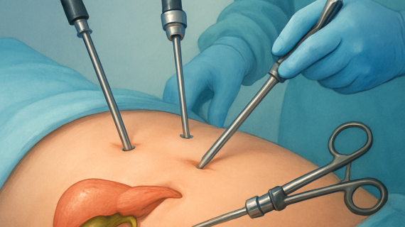
Laparoscopic or Open Cyst Fenestration/Aspiration
Cyst fenestration, also known as deroofing, involves surgically opening the cyst, excising a portion of its wall, and allowing the remaining cyst lining to collapse and adhere to the liver parenchyma. This can be performed laparoscopically or via open surgery and is effective in relieving symptoms from large simple cysts. Simple aspiration of cyst fluid is generally not curative as the cyst will likely reform.
In conclusion, the assessment and management of liver cysts form a nuanced clinical challenge. A systematic approach, beginning with accurate classification and leveraging the power of modern imaging modalities, is essential. While simple cysts often necessitate a period of observation, complex lesions and those with specific etiologies demand tailored treatment strategies, ranging from targeted antifungal or antibiotic therapy and minimally invasive techniques to definitive surgical resection. The ultimate goal is to ensure patient well-being by accurately diagnosing the nature of the cyst and implementing the most appropriate management plan, thus transforming a potential clinical puzzle into a well-managed outcome.
FAQs
What is a liver cyst and how is it classified?
A liver cyst is a fluid-filled sac that forms in the liver. Liver cysts can be classified into simple cysts, which are benign and usually asymptomatic, and complex cysts, which may have septations, calcifications, or solid components and require further evaluation. Other classifications include parasitic cysts (such as hydatid cysts) and cystic neoplasms.
How are liver cysts diagnosed using imaging techniques?
Liver cysts are primarily diagnosed through imaging modalities such as ultrasound, computed tomography (CT), and magnetic resonance imaging (MRI). Ultrasound is often the first-line tool to detect cysts, while CT and MRI provide detailed characterization to differentiate simple cysts from complex or potentially malignant lesions.
When is surveillance recommended for liver cysts?
Surveillance is generally recommended for simple liver cysts that are asymptomatic and have benign imaging features. Periodic imaging follow-up may be advised to monitor for changes in size or characteristics, especially if the cyst is large or has atypical features.
What are the management options for symptomatic or complicated liver cysts?
Management options for symptomatic or complicated liver cysts include percutaneous aspiration, sclerotherapy, and surgical intervention such as cyst fenestration or resection. The choice of treatment depends on the cyst’s size, symptoms, risk of complications, and underlying pathology.
Can liver cysts become cancerous?
Simple liver cysts are typically benign and do not become cancerous. However, certain complex cystic lesions or cystic neoplasms have a potential for malignancy and require careful evaluation and management to rule out or treat cancerous changes.