We will begin by dissecting the anatomy of the duodenum, that crucial initial segment of our small intestine. This is not merely a passive tube, but a dynamic chamber where the symphony of digestion truly begins in earnest. Imagine it as the grand reception hall of the digestive tract, where guests—the food we consume—are met with a cascade of essential tools to break them down. Its structure, blood supply, and the clinical ramifications of its well-being are vital for understanding our overall health.
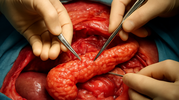
The duodenum, the very first part of our small intestine, is a remarkably compact yet profoundly important organ. Spanning approximately 23 to 30 centimeters, it is the shortest segment of this vital organ system. Its characteristic C- or horseshoe-shape cradles the head of the pancreas, a close anatomical relationship that is central to its function. We can conceptually divide this structure into four distinct parts, each with its own anatomical nuances and functional significance.
The Four Parts: A Sequential Journey Through the Upper Small Intestine
We need to understand these four segments as a continuous pathway, each playing its role in ushering our partially digested food from the stomach onward.
The Superior Part: The Initial Reception
The superior part of the duodenum is the shortest of the four, measuring around 5 centimeters in length. It commences immediately after the pyloric sphincter, the muscular valve that controls the passage of food from the stomach into the duodenum. This initial segment is often referred to as the duodenal bulb or cap. At its very beginning, it is an intraperitoneal structure, meaning it is suspended within the peritoneal cavity by a mesentery. However, as it progresses, it transitions to become retroperitoneal, lying behind the peritoneum. This initial region is critical because it is here that the acidic chyme from the stomach is first buffered and begins to be mixed with digestive juices. We will later see how this region is particularly susceptible to certain digestive challenges.
The Descending Part: The Ductial Crossroads
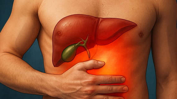
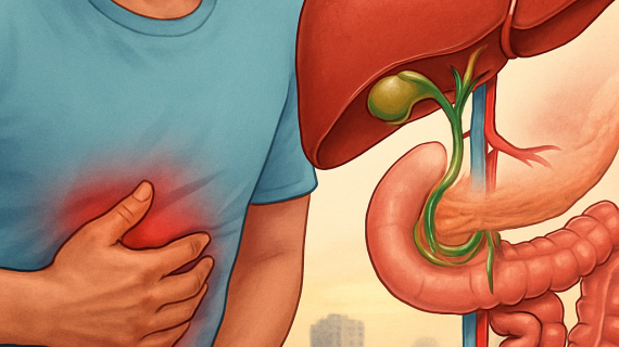
Following the superior part is the descending or second part of the duodenum, which is considerably longer, typically measuring between 10 to 12 centimeters. This segment takes a sharp turn downwards, following the curve of the pancreatic head. Its most significant feature is its role as the point of entry for essential digestive secretions from two vital accessory organs: the liver and the pancreas. The common bile duct, carrying bile produced by the liver, and the main pancreatic duct, carrying enzymes from the pancreas, converge to empty their contents into the duodenum. This convergence typically occurs at a small elevation within the duodenal wall known as the major duodenal papilla, or the ampulla of Vater. This papilla is a focal point where the chemical breakdown of our food truly intensifies, facilitated by the precise timing and delivery of bile and pancreatic enzymes.
The Horizontal/Inferior Part: Traversing the Vascular Highway
The horizontal or third part of the duodenum extends approximately another 10 to 12 centimeters, running transversely across the abdomen from right to left. It passes anteriorly to the aorta and the inferior vena cava, two major blood vessels that form the central vascular highway of our body. This anatomical position is significant because it places the duodenum in close proximity to these critical vessels. We will explore later how this relationship can have significant clinical implications, particularly in certain conditions that affect the arteries supplying the intestines.
The Ascending Part: The Final Turn and Transition
The final segment is the ascending or fourth part of the duodenum, which is the shortest, measuring only about 2 to 3 centimeters. It courses upwards from the horizontal part and then makes a sharp bend posteriorly. This bend marks the transition from the duodenum to the next section of the small intestine, the jejunum. This crucial junction is facilitated by a strong band of muscle and connective tissue known as the ligament of Treitz. The ligament of Treitz is not just a simple anatomical landmark; it serves as a critical suspensory muscle that helps to maintain the position of the duodenojejunal flexure and plays a role in preventing kinks or obstructions at this vital juncture.
In addition to understanding the intricate anatomy of the duodenum, readers may find it beneficial to explore the related article on the art of engaging readers through compelling titles. This resource provides valuable insights into how effective communication can enhance the presentation of complex topics, such as the detailed structure and clinical importance of the duodenum. For more information, you can visit the article here: The Art of Drawing Readers In.
The Microscopic Architecture: The Layers That Build the Duodenum
Beyond its macroscopic structure, the duodenum, like all hollow organs in our body, is built from distinct layers, each contributing to its overall function. We can liken these layers to the different tradespeople involved in constructing a sophisticated building, each with a specialized role. These layers are the mucosa, submucosa, muscularis, and serosa.
The Four Fundamental Layers: A Closer Look
Understanding this microscopic anatomy is key to appreciating how the duodenum performs its complex tasks.
The Mucosa: The Inner Lining and Digestive Hub
The innermost layer, the mucosa, is where the magic of digestion and absorption truly happens. It is lined with simple columnar epithelium, a single layer of tall, rectangular cells. These cells are equipped with countless microscopic projections called microvilli, which collectively form a brush border. This brush border dramatically increases the surface area available for nutrient absorption, much like adding more shelves to an already expansive library to hold more books. Within the mucous membrane itself, we find specialized glands. Most notably, in the duodenum, we encounter Brunner’s glands. These are compound tubular glands located within the submucosa, but their ducts open into the crypts of Lieberkühn, which are invaginations of the surface epithelium. Brunner’s glands are unique to the duodenum and secrete an alkaline mucus that is crucial for neutralizing the acidic chyme entering from the stomach, protecting the duodenal lining and creating an optimal pH for the action of pancreatic enzymes.
The Submucosa: The Support System
Beneath the mucosa lies the submucosa, a layer of dense connective tissue. This layer is rich in blood vessels and lymphatic vessels, which are essential for transporting absorbed nutrients. It also contains nerves that help regulate the muscular activity and secretions of the duodenum. The submucosa provides structural support for the mucosa and plays a vital role in the overall nourishment and function of the duodenal wall.
The Muscularis: The Engine of Movement
The muscularis layer is responsible for the peristaltic movements of the duodenum, the wave-like contractions that propel food forward. This layer consists of two sub-layers of smooth muscle: an inner circular layer and an outer longitudinal layer. The coordinated contraction and relaxation of these muscle fibers churn the food, mix it thoroughly with digestive juices, and move it along the intestinal tract. Without this muscular engine, our food would simply stagnate.
The Serosa: The Protective Outer Coat
The outermost layer of the duodenum is the serosa, which is a thin, smooth membrane. In the intraperitoneal portions of the duodenum, this serosa is a part of the visceral peritoneum. However, in the retroperitoneal portions (which constitute the majority), the serosa is either absent or very thin, as the organ is directly adherent to the posterior abdominal wall. This outer layer provides a protective covering and helps to reduce friction between organs within the abdominal cavity.
The Duodenum’s Lifeline: Blood Supply and Drainage

The duodenum, like any metabolically active tissue, requires a robust blood supply to deliver oxygen and nutrients and to remove waste products. Its arterial supply is a fascinating interplay of vessels originating from different embryological regions, reflecting its complex development.
Arterial Supply: A Dual Origin
We can visualize the arterial supply of the duodenum as two major rivers feeding into a network of smaller streams. This ensures that this vital organ is well-irrigated.
The Superior Pancreaticoduodenal Arteries: The Foregut’s Contribution
The superior pancreaticoduodenal arteries are responsible for irrigating the initial segments of the duodenum, including the bulb and the descending portion. These arteries are branches of the gastroduodenal artery, which itself is a branch of the common hepatic artery. The common hepatic artery, as we know, originates from the celiac trunk, the first major arterial branch off the abdominal aorta, which supplies the foregut derivatives. These arteries provide oxygenated blood and nutrients essential for the intense digestive and absorptive processes occurring in this region.
The Inferior Pancreaticoduodenal Arteries: The Midgut’s Support
The inferior pancreaticoduodenal arteries supply the latter parts of the duodenum, namely the horizontal and ascending portions, and also contribute to the blood supply of the pancreas. These arteries originate from the superior mesenteric artery. The superior mesenteric artery is the primary artery supplying the midgut derivatives. This dual arterial supply, originating from both the foregut and midgut arterial systems, highlights the embryological origin of the duodenum, which is formed from the fusion of foregut and midgut tissues. These arteries branch and anastomose, creating a rich vascular network ensuring adequate perfusion.
Venous Drainage: The Return Journey
The venous drainage of the duodenum mirrors its arterial supply. The blood from the superior pancreaticoduodenal veins typically drains into the gastroduodenal vein, which then contributes to the portal venous system. Similarly, the blood from the inferior pancreaticoduodenal veins drains into the superior mesenteric vein, which also forms a major component of the portal vein. Ultimately, the venous blood from the duodenum, along with blood from other abdominal digestive organs, collects in the portal vein and is transported to the liver for processing before returning to the general circulation. This coordinated arterial supply and venous drainage ensures the efficient removal of metabolic byproducts and the delivery of essential substances.
For reliable health advice, Get best medical expert consultation today.
The Clinical Significance: Where Anatomy Meets Health and Disease
The duodenum, due to its unique structure, location, and crucial role in digestion, is a frequent site of various clinical conditions. Understanding its anatomy is paramount for diagnosing and managing these issues.
For those interested in a deeper understanding of gastrointestinal anatomy, the article on Duodenum Anatomy Explained With Detailed Structure, Parts, Blood Supply, and Clinical Importance provides an extensive overview. This resource not only covers the intricate details of the duodenum but also highlights its crucial role in digestion and nutrient absorption. Exploring related topics can enhance your comprehension of how the digestive system functions as a whole, making it essential for both students and healthcare professionals.
Common Duodenal Pathologies: Vulnerabilities of the First Section
We must recognize that even the most crucial organs can be susceptible to illness.
Peptic Ulcers: The Scars of Acidity
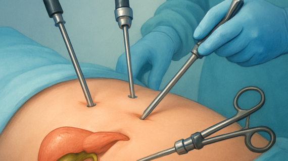
The most common clinical issue associated with the duodenum is the development of peptic ulcers. These are open sores that form on the lining of the digestive tract. The duodenal bulb, the initial part of the superior duodenum, is particularly prone to ulcer formation. This susceptibility is largely due to the direct exposure to highly acidic chyme leaving the stomach. While the Brunner’s glands provide some alkaline buffering, excessive acid production or a weakened mucosal defense can lead to ulceration. The bacteria Helicobacter pylori is a major contributor to duodenal ulcers by damaging the protective mucous layer and increasing acid production. The presence of these ulcers can manifest as burning abdominal pain, often relieved by eating or antacids, and in severe cases, can lead to bleeding or perforation.
The Digestive Powerhouse: Role in Nutrient Assimilation
The duodenum is not just a conduit; it is a critical site for the initial stages of digestion and absorption. It receives pancreatic enzymes that break down carbohydrates, proteins, and fats into smaller molecules. Bile, arriving from the liver and gallbladder, aids in fat digestion by emulsification. Furthermore, the duodenum plays a vital role in the absorption of specific nutrients. It is the primary site for the absorption of iron, essential for our red blood cells, and calcium, crucial for bone health. Any disruption to the duodenum’s structure or function can therefore have cascading effects on our body’s ability to extract essential nutrients from our food.
Anatomical Relationships and Potential Complications: A Delicate Proximity
The close anatomical relationship of the duodenum with vital organs like the pancreas, liver, kidneys, and major blood vessels like the aorta and inferior vena cava can lead to significant clinical problems. We can think of this proximity as a crowded neighborhood where proximity can sometimes lead to unexpected pressures. For instance, conditions that cause swelling or displacement of the pancreas can compress the duodenum.
Superior Mesenteric Artery (SMA) Syndrome: Avascular Squeeze
A particularly noteworthy example is Superior Mesenteric Artery (SMA) syndrome. Normally, the superior mesenteric artery passes anterior to the horizontal (third) part of the duodenum. However, in certain individuals, particularly those who experience rapid weight loss or have an unusually mobile duodenum, the angle between the aorta and the SMA can become narrowed. This can lead to the SMA compressing the duodenum between itself and the aorta, obstructing the passage of food. This syndrome can present with symptoms similar to gastric outlet obstruction, including nausea, vomiting, and abdominal pain, especially after meals.
Embryological Origins and Fixed Positions: The Scars of Development
The embryological development of the duodenum is a fascinating narrative. It arises from both the foregut and the midgut. As the embryonic gut tube develops and rotates, the duodenum forms and becomes largely fixed in its retroperitoneal position due to the growth and adherence of the liver and pancreas to the posterior abdominal wall. This fixation, while essential for maintaining its position, also contributes to its vulnerability. If surrounding structures shift or swell, the fixed duodenum can be impinged upon. Understanding these developmental origins helps explain why certain congenital anomalies, though rare, can affect the duodenum.
In conclusion, the duodenum is far more than just the first few inches of our small intestine. It is a meticulously designed segment, a crucible where digestive processes are initiated with remarkable efficiency, a critical interface for nutrient absorption, and a structure whose anatomical relationships and intrinsic vulnerabilities make it a focal point for numerous clinical concerns. Our understanding of its intricate anatomy, from the microscopic brush border to its arterial lifeline and its proximate neighbors, is fundamental to appreciating the delicate balance of our digestive health.
FAQs
What is the duodenum and where is it located?
The duodenum is the first section of the small intestine, located immediately after the stomach. It connects the stomach to the jejunum and plays a crucial role in digestion by receiving partially digested food and mixing it with digestive enzymes.
What are the main parts of the duodenum?
The duodenum is divided into four parts: the superior (first) part, descending (second) part, horizontal (third) part, and ascending (fourth) part. Each part has specific anatomical features and relationships with surrounding organs.
How is the duodenum supplied with blood?
The duodenum receives blood from branches of both the celiac trunk and the superior mesenteric artery. The superior pancreaticoduodenal artery (from the gastroduodenal artery) and the inferior pancreaticoduodenal artery (from the superior mesenteric artery) are the primary vessels supplying the duodenum.
What is the clinical importance of the duodenum?
The duodenum is clinically significant because it is a common site for peptic ulcers, which can cause pain and bleeding. It also plays a role in nutrient absorption and is involved in conditions such as duodenal obstruction and tumors.
How does the duodenum contribute to digestion?
The duodenum receives chyme from the stomach and mixes it with bile from the liver and digestive enzymes from the pancreas. This process helps neutralize stomach acid and breaks down nutrients, facilitating their absorption in the small intestine.