If you live in Dhaka and are facing upper abdominal pain, jaundice, recurrent gallbladder attacks or unexplained abnormal blood tests, early recognition of liver health problems changes what treatment is possible. This practical primer groups the warning patterns to watch, explains which blood tests and imaging truly guide decisions, and gives clear thresholds for urgent versus outpatient referral to a hepatobiliary surgeon at Popular Medical College Hospital, Dhanmondi. It finishes with a concise checklist for your first visit so you can move from uncertainty to a focused plan.
1. Why liver and biliary health matters in Dhaka
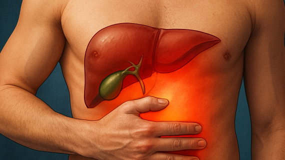
Direct point: Liver health in Dhaka affects whether a common symptom like upper abdominal discomfort remains a simple outpatient problem or becomes a time-sensitive surgical emergency. Problems that start as gallbladder pain, fatty liver, or mild jaundice can progress to obstructive jaundice, sepsis, or unresectable tumours if diagnosis and referral are delayed.
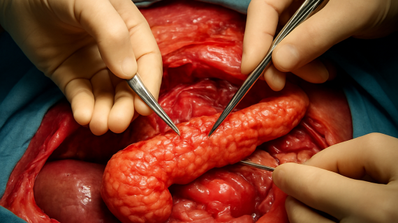
Local pattern: In practice I see three frequent drivers here – symptomatic gallstones, rising rates of nonalcoholic fatty liver disease linked to diabetes and sedentary life, and a persistent burden of viral hepatitis leading to cirrhosis and hepatocellular carcinoma. Pancreatic head tumours and extrahepatic bile duct cancers are less common but present late and require coordinated surgical care. For background on viral hepatitis, see WHO on hepatitis.
Important trade-off: Early imaging and specialist review preserve curative options but cost and access matter. Ultrasound is widely available and usually sufficient to detect gallstones or bile duct dilation; CT and MRI with MRCP offer staging detail but are not necessary for every patient. Choosing when to escalate from ultrasound to cross-sectional imaging is a clinical judgement that balances urgency, cost, and how the result will change management.
Role of surgical judgement: A hepatobiliary surgeon does more than operate. We decide timing for ERCP versus surgery, assess resectability of liver tumours, and coordinate preoperative optimization for patients with poor liver function. In my experience early surgical input often prevents a cascade of avoidable interventions and keeps curative pathways open, especially for obstructive jaundice and resectable hepatic lesions. For local services see services.
Concrete example: A 52 year old man from Dhanmondi developed progressive jaundice over two weeks. Ultrasound showed dilated intrahepatic ducts but no clear gallstones in the gallbladder. He underwent ERCP for biliary decompression, CT staging, and then hepatic resection at Popular Medical College Hospital after multidisciplinary review. Early decompression and timely staging changed a potentially palliative situation into a curative plan.
Common misconception and practical advice: Many people assume normal general health means the liver is fine. That is false – fatty liver can be silent and bile duct obstruction can present with predominantly itching or dark urine before severe pain. Bring any imaging reports and recent liver function tests to your first specialist visit, and avoid unprescribed herbal cleanses or alcohol which commonly worsen liver inflammation.
2. Early symptom clusters to watch for
Direct point: Patterns of symptoms, not isolated complaints, determine how quickly you need investigation and which tests will be useful. Recognising one of the following clusters helps your clinician decide between urgent decompression, expedited imaging, or routine outpatient workup.
A decision-oriented symptom framework
| Cluster | Key symptoms and timing | Likely causes | Action that changes outcomes |
|---|---|---|---|
| Biliary colic | Episodic right upper quadrant pain, often after fatty meals, with nausea; pain settles between attacks | Gallstones in the gallbladder | Plan elective laparoscopic cholecystectomy when attacks are recurrent; pain-only cases can wait for outpatient surgical review |
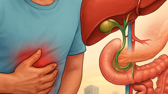
| Cholestatic obstruction | Progressive yellowing of the eyes/skin, dark urine, pale stools, itching over days to weeks | Common bile duct stone, stricture, or tumour obstructing bile flow | Urgent imaging and likely biliary decompression (ERCP or percutaneous drainage) if bilirubin is rising |
| Hepatocellular dysfunction | Persistent fatigue, abdominal fullness, easy bruising or new abdominal swelling over weeks to months | Fatty liver, chronic viral hepatitis, cirrhosis or liver tumour | Blood panel and targeted imaging for staging or medical management; surgical options depend on reserve and lesion resectability |
| Infective/inflammatory | High fevers with rigors, severe continuous RUQ pain, hypotension or confusion | Ascending cholangitis or infected bile collection | Emergency admission and urgent biliary drainage within 24 hours |
Practical limitation: Normal or mildly abnormal liver enzymes do not exclude significant biliary obstruction or an evolving liver tumour. Enzyme patterns lag, and ultrasound is operator dependent. That means clinical pattern and trend matter more than a single test result.
Tradeoff to understand: MRCP maps biliary anatomy without risk and guides surgery, but it does not relieve obstruction. ERCP treats obstruction immediately yet carries procedural risk. In practice reserve ERCP for patients who are symptomatic with biochemical cholestasis or sepsis, and use MRCP for planning when urgency is lower.
Concrete Example: A 48 year old woman had three short episodes of severe postprandial RUQ pain over two months and was treated for gastritis. On the fourth episode she developed jaundice; ultrasound showed dilated bile ducts. Timely referral led to ERCP stone extraction and a planned laparoscopic cholecystectomy, avoiding recurrent obstruction and pancreatitis.
- Watch for atypical signals: intense generalized itching or pale stools often precede obvious jaundice and point to cholestasis rather than simple gastritis.
- Nonpainful jaundice is real: tumours can obstruct bile ducts with little pain, so absence of severe pain does not reassure.
- New metabolic changes: unexplained weight loss or new onset diabetes with upper abdominal symptoms increases suspicion for pancreatic head pathology and should shorten the diagnostic timeline.
Takeaway: Match the symptom cluster to urgency: episodic pain usually allows planned surgical care, cholestasis and infection demand faster imaging and possible biliary drainage. Prepare by bringing symptoms timeline, recent LFTs, and any ultrasound when you come for review.
3. Which initial tests matter and why
Short answer: focus on a small set of tests that actually change decisions: liver function panel (pattern, not a single value), full blood count/CRP, targeted viral and metabolic tests, abdominal ultrasound, and selective use of AFP or CA 19-9 and cross‑sectional imaging when the first-line results point to tumour or obstruction.
Interpreting results the way a surgeon needs them
Pattern over single numbers: a cholestatic pattern (high bilirubin, alkaline phosphatase, GGT) moves you toward urgent biliary imaging and possible decompression; a hepatocellular pattern (high ALT/AST) prompts metabolic, viral and fatty liver evaluation and staged imaging. Trends are more useful than one-off abnormalities.
- Liver function tests (LFTs): bilirubin, ALT, AST, alkaline phosphatase, GGT, albumin and INR — these classify obstruction versus parenchymal disease and indicate synthetic reserve.
- Full blood count and CRP: essential if infection or cholangitis is suspected; white cell count and CRP guide urgency.
- Viral serology and metabolic screen: HBsAg, anti-HCV, fasting glucose and lipid profile when NAFLD or viral hepatitis is possible; important for long-term management and surgical risk.
- Abdominal ultrasound: first-line imaging for gallstones, biliary dilation and obvious liver lesions; cheap and available but operator dependent.
- Tumour markers: AFP for patients with cirrhosis or chronic HBV at risk of hepatocellular carcinoma; CA 19-9 only useful alongside suspicious imaging for pancreaticobiliary disease — both have limited sensitivity and produce false positives.
Key tradeoff: ultrasound gives quick, actionable information for most patients and should usually come before expensive MRCP or CT. Reserve MRCP/contrast CT when ultrasound is inconclusive or when imaging will change an operative plan. ERCP treats obstruction but is not a diagnostic first step unless decompression is immediately needed.
Clinical illustration: a patient has mildly elevated ALT and ultrasound showing a 2 cm solitary liver lesion. Ordering AFP and a contrast MRI clarified the lesion as likely hepatocellular carcinoma, allowing timely referral for resection planning at Popular Medical College Hospital rather than months of serial ultrasound that would delay curative surgery.
Practical limitation and judgment: tumour markers are overused as screening tools. In practice, AFP helps in high‑risk surveillance but a normal AFP does not rule out cancer. Likewise, minor enzyme abnormalities often reflect fatty liver and lifestyle factors — treatable medically — yet rising bilirubin or ductal dilation demands faster intervention.
Match the biochemical pattern to the imaging test: cholestasis → urgent biliary imaging/decompression; hepatocellular pattern → viral/metabolic workup and targeted cross‑sectional imaging.
4. Imaging and endoscopic tests: when to use ultrasound, CT, MRI, MRCP and ERCP
Direct guidance: Start with abdominal ultrasound for almost every patient with suspected biliary pain, jaundice or abnormal liver function tests. Ultrasound detects gallstones, shows bile duct dilation and is cheap and widely available in Dhaka. If ultrasound is normal but clinical suspicion remains high, choose the next test based on whether you need treatment now or anatomic detail for planning.
How each modality changes decisions
CT (contrast enhanced multiphase): use CT when you need staging, vascular mapping or to evaluate a suspected hepatic or pancreatic mass. CT is faster than MRI and excellent for surgical planning of larger tumours, but it exposes the patient to radiation and iodinated contrast so avoid it as a routine follow up for simple gallstones.
MRI and MRCP: choose MRI with MRCP when you need high soft tissue contrast or noninvasive biliary anatomy. MRCP picks up small intrahepatic strictures and clarifies equivocal ultrasound findings without the risks of ERCP. Limiting factors are cost, scanner availability and contraindications such as certain implants or severe claustrophobia.
ERCP: reserve ERCP primarily as a therapeutic procedure for biliary decompression, stone extraction and stent placement. ERCP treats obstruction but carries measurable risk of pancreatitis and infection. Do not use ERCP as the first diagnostic step unless the patient needs urgent drainage or you plan immediate intervention.
Practical consideration and tradeoff: pick the least invasive test that will change management. MRCP often looks prettier but will not relieve obstruction; ERCP fixes the problem but has procedural risk. In patients who are septic or have rapidly rising bilirubin, prioritise urgent ERCP or percutaneous drainage over waiting for MRCP.
- Typical pathway for obstructive cholestasis: ultrasound first, then urgent ERCP if signs of sepsis or rising bilirubin; if stable and diagnosis unclear, MRCP followed by targeted CT or MRI for staging.
- Typical pathway for a solitary liver lesion on ultrasound: contrast MRI for lesion characterisation; reserve CT when you need vascular mapping for resection.
- When ultrasound is limited: in obese patients or technically poor scans, repeat ultrasound at a specialist centre or proceed to MR imaging rather than relying on a single poor study.
Real-world case: A 60 year old woman presented with cholestatic LFTs and mild jaundice. Ultrasound showed intrahepatic duct dilation but no clear stone. Because she was clinically stable we performed MRCP the same day, identified a distal bile duct stricture, and scheduled ERCP for targeted stenting the next morning followed by CT staging. This sequence avoided unnecessary blind ERCP and provided a clear surgical plan.
Judgment call surgeons make: Many clinicians reflexively order both CT and MRCP. That duplicates cost and delays care. In my practice I ask one question: will this test change what I do in the next 48 hours? If not, defer the higher cost study and use focused imaging only when it meaningfully alters the operative or endoscopic plan. For local coordination see services and for general guidance on gallstones see NHS on gallstones.
5. Clear referral criteria: when to see a hepatobiliary surgeon immediately and when to arrange expedited outpatient review
Direct rule: refer immediately when there is clinical or biochemical evidence that bile flow is blocked and infection or systemic compromise is present. Waiting for a clinic appointment in these cases removes options and increases risk.
Immediate (within hours): any patient with the combination of fever or rigors, jaundice, and new severe right upper quadrant pain – or obvious sepsis with cholestatic changes on bloods – needs emergency assessment and biliary drainage. Look for a rising bilirubin over serial samples, hypotension, altered mental state, or marked leukocytosis. In practice this means admission, urgent imaging and usually ERCP or percutaneous drainage before definitive surgery.
Expedited outpatient review (days to 2 weeks): patients who are stable but have recurrent symptomatic gallstone attacks, a documented episode of biliary pancreatitis, or imaging showing bile duct dilation or a solitary suspicious liver lesion should see a hepatobiliary surgeon quickly. These cases often require timed ERCP, MRCP or contrast imaging plus definitive planning – delay beyond two weeks commonly leads to repeat admissions or loss of resectability for tumours.
Routine surgical assessment (4 weeks or as appropriate): isolated, mild enzyme abnormalities without symptoms, or known fatty liver with stable tests, can be worked up in the outpatient setting with lifestyle and medical management first. Use specialist referral when imaging or test trends change, or when synthetic function is impaired.
Key practical tradeoff: decompress first, operate later. Treating active cholangitis with ERCP or drainage reduces operative risk and expands surgical options. Conversely, taking a febrile, jaundiced patient straight to major hepatic resection without decompression increases complications. The surgeon must balance urgency against physiologic optimization – this is not theoretical, it matters in outcomes.
Judgment call many clinicians miss: a single normal ultrasound or mildly abnormal ALT does not exclude obstructive disease or significant liver pathology. Trends, symptom pattern, and the presence of systemic signs should drive referral more than a solitary test. In Dhaka resource constraints sometimes delay MRCP; when MRCP is unavailable, plan management around the test that will change treatment in the next 48 hours.
Concrete example: A 55 year old woman had two admissions for biliary pancreatitis in six weeks. After stabilisation and a same admission ERCP stone clearance she was scheduled for laparoscopic cholecystectomy within 10 days to prevent recurrence. Early surgical scheduling prevented a third admission and simplified definitive care.
- What to bring to a referral: recent LFTs showing trends, ultrasound or CT reports, notes from any ERCP admission and a clear timeline of symptoms.
- When you will be admitted: fever plus jaundice plus pain, rapid bilirubin rise, hypotension or confusion.
- When outpatient is acceptable: recurrent colic without systemic features, stable solitary small lesions requiring characterisation, or mild enzyme abnormalities under investigation.
If you have fever plus jaundice plus severe pain, seek emergency care. For prompt specialist planning and to avoid repeated admissions bring imaging and serial LFTs to your appointment.
6. What to expect at a hepatobiliary surgical consultation and common surgical options
Direct point: the first hepatobiliary surgical visit is an active decision session, not just paperwork. Expect focused review of your symptoms, prior scans and blood tests, and a clear plan that either schedules definitive treatment or orders targeted investigations that will change the immediate plan.
What the consultation covers
History and risk assessment: the surgeon will clarify timing and pattern of pain or jaundice, alcohol and viral hepatitis history, metabolic risks for fatty liver, prior operations and current medications. Imaging reconciliation: bring your ultrasound, CT or MRI; the surgeon compares reports to decide if further imaging will change the operation. Functional assessment: liver function, coagulation and basic cardiopulmonary fitness are reviewed because they determine whether a planned resection is safe.
Common options you will discuss in plain language
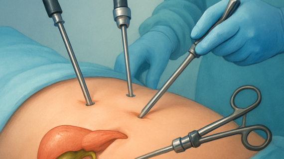
Plan descriptions are concrete. For symptomatic gallstones the usual option is laparoscopic cholecystectomy (small wounds, quick return). For stones or blockages in the bile duct expect either ERCP with stone extraction or stent (endoscopic decompression) or a direct common bile duct exploration during surgery. For a liver tumour the discussion shifts to the type and extent of hepatic resection (removing a segment or lobe) with attention to how much healthy liver must remain. Pancreatic head tumours may require a pancreaticoduodenectomy (Whipple) — major but potentially curative when staged appropriately.
Trade-offs and limits: minimally invasive approaches reduce pain and hospital stay but are not always appropriate. Inflamed tissue, recent infection, prior surgery or complex vascular anatomy push a surgeon to choose open surgery for safety. Also, obstructed patients often need decompression first (ERCP or percutaneous drainage) because operating on a jaundiced, septic patient raises complication risk.
Preoperative optimisation matters: expect requests for nutritional support, diabetes control, vaccination for hepatitis if relevant, and sometimes a referral to anaesthesia for cardiopulmonary assessment. For major liver resection we check liver function and calculate future liver remnant — if reserve is marginal we delay or plan staged procedures.
Concrete example: a 58 year old man with obstructive jaundice had ERCP stenting on the day of consultation to lower bilirubin and treat cholangitis. Two weeks of optimisation followed, then CT mapping and elective right hemihepatectomy with an uneventful recovery — decompression first preserved the option for curative resection.
7. Practical next steps for patients in Dhaka and how to arrange care with Dr Murshidul Arefin
Start with triage, not appointment anxiety. First decide whether your problem is urgent (fever plus jaundice, severe unrelieved pain, rapid bilirubin rise) or suitable for expedited outpatient review. That decision determines the pathway: emergency admission and possible ERCP the same day, or a planned clinic visit with targeted imaging and multidisciplinary planning.
Step-by-step actions to get the right care quickly
- Classify urgency: If you have fever with jaundice or worsening confusion, go to the nearest emergency department and tell the staff you need hepatobiliary assessment. For nonseptic but progressive jaundice or recurrent biliary pain, request an expedited outpatient slot within 48–72 hours.
- Prepare your records: Assemble lab reports with dates and reference ranges (LFTs, INR), and radiology files. Prefer digital DICOMs or CDs so the surgical team can re-review imaging; printed reports alone are often insufficient for operative planning.
- Book the right appointment: Use the clinic contact to request either an urgent hepatobiliary review or a routine consult. For services and booking use services or contact — specify symptom timeline and whether you were admitted recently for ERCP or pancreatitis.
- Expect a focused triage call: The team will tell you whether to bring original test results, whether repeat bloods or an urgent ultrasound/MRCP is needed, and whether admission will be required for endoscopic drainage before surgery.
- Coordinate logistics: If surgery is likely, arrange baseline optimisation: diabetic control, nutrition, vaccination status for hepatitis if applicable, and any necessary cardiopulmonary clearance. Ask the clinic about estimated costs and whether preauthorisation is needed for insurance.
- Clarify follow up and contingency: If imaging is pending locally, request secure transfer of images to Popular Medical College Hospital, Dhanmondi to avoid repeated scans. If symptoms escalate while waiting for an appointment, present to emergency care and mention your scheduled consultation.
Practical tradeoff: A teleconsult is useful for triage and reducing needless travel, but it cannot replace hands-on assessment when obstruction or infection is suspected. Expect telemedicine to set the plan; expect in-person visits for endoscopy, same-day ERCP scheduling, and operative consent.
Concrete example: A patient from Mirpur phoned for an urgent review after two weeks of worsening jaundice but no fever. The clinic arranged an expedited MRCP and a same-week slot with Dr Murshidul Arefin. MRCP showed a distal bile duct stricture; she underwent ERCP stenting the next day, followed by multidisciplinary staging and elective resection three weeks later. The staged approach avoided emergency surgery and improved operative planning.
Bring: digital imaging (DICOM or secure link), LFTs with dates, list of medications and a clear timeline of symptoms — this saves time and prevents repeated tests.
One judgment many patients miss: focusing on liver supplements or a quick liver detox delays meaningful care when structural problems are present. For suspected fatty liver or metabolic causes, expect medical management (diet, exercise, diabetes control, statin for cholesterol when indicated) rather than surgery. For obstructive patterns, procedural decompression and targeted imaging matter far more than any cleanse or supplement.